Hip Muscles Diagram Labeled : Hip Thigh Atlas Of Anatomy. Hip pain on the outside of your hip, upper thigh or outer buttock is usually caused by problems with muscles, ligaments, tendons and other soft tissues that surround your hip joint. Human muscle system, the muscles of the human body that work the skeletal system, that are under voluntary control, and that are concerned with movement, posture, and balance. The thigh bone or femur and the pelvis join to form the hip joint. In general, they work in pairs. The iliopsoas actually consists of two muscles:
Hip pain on the outside of your hip, upper thigh or outer buttock is usually caused by problems with muscles, ligaments, tendons and other soft tissues that surround your hip joint. It allows us to walk, run, and jump. See more ideas about muscle diagram, medical anatomy, body anatomy. These muscles can be grouped based upon their location and function. It is also referred to as a ball and socket joint and is surrounded by muscles, ligaments, and tendons.
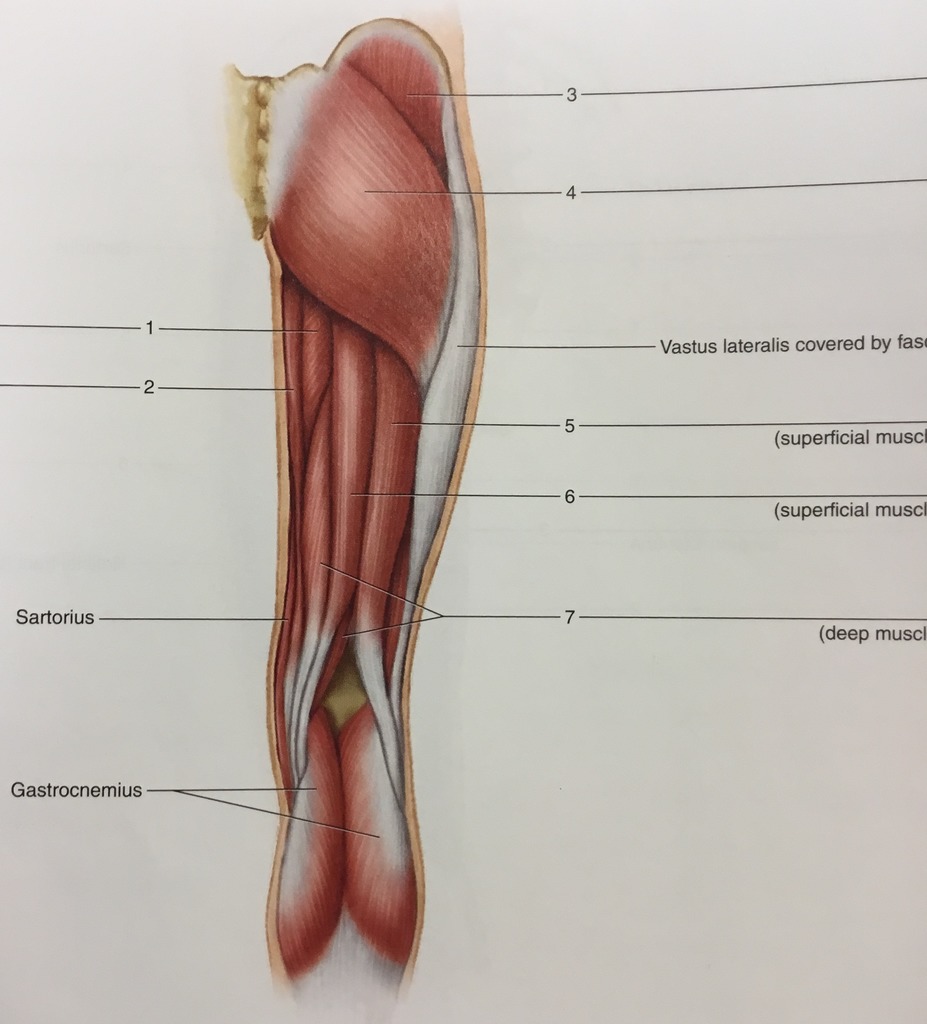
Muscle Lab 23 Figure 23 3 Muscles Of The Posterior Right Hip And Thigh Diagram Quizlet from o.quizlet.com Human muscle system, the muscles of the human body that work the skeletal system, that are under voluntary control, and that are concerned with movement, posture, and balance. The hip muscles encompass many muscles of the hip and thigh whose main function is to act on the thigh at the hip joint and stabilize the pelvis.without them, walking would be impossible. Any injury or disease of the hip will adversely affect the joint's range of motion and ability to bear weight.</p> There are two hip bones, one on the left side of the body and the other on the right. Muscles of thigh and the hip anatomy the pelvic girdle and the vertebral column both yield the various muscles responsible for initiating movement of the thigh at the level of the hip. The hip muscles are composed of multiple flexors, extensors, adductors, abductors, and rotators that work together. The superficial muscles of the thigh. The hip joint is one of the most flexible joints in the entire human body.
Learn vocabulary, terms, and more with flashcards, games, and other study tools.
They also stabilise the hip joint by 'pulling' the femoral head into the acetabulum of the pelvis. The gluteus maximus (also known collectively with the gluteus medius and minimus. Click on the labels below to find out more about your muscles. They can be divided into three main groups: The bones together make up the hip. See more ideas about muscle diagram, medical anatomy, body anatomy. The iliopsoas actually consists of two muscles: Hip muscle anatomy is a complex topic. The hip muscles are composed of multiple flexors, extensors, adductors, abductors, and rotators that work together. These muscles are not only vital in the process of creating movement, but they are also vital in providing stability. Muscles of thigh and the hip anatomy the pelvic girdle and the vertebral column both yield the various muscles responsible for initiating movement of the thigh at the level of the hip. The hip flexors are several muscles that bring your legs and trunk together in a flexion movement. We are pleased to provide you with the picture named muscles of hip and thigh lateral view.we hope this picture muscles of hip and thigh lateral view can help you study and research.
Anatomynote.com found muscles of hip and thigh lateral view from plenty of anatomical pictures on the internet. The femur may also rotate around its axis about 90 degrees at the hip. These muscles then attach at their points of insertion along the femur at various locations. We have a lot of muscles in our bodies (literally, over 600). They also stabilise the hip joint by 'pulling' the femoral head into the acetabulum of the pelvis.
3 from For more anatomy content please follow us and visit our website: Anatomy of the hip muscles. It is also referred to as a ball and socket joint and is surrounded by muscles, ligaments, and tendons. Muscles allow us to move and function. There are two hip bones, one on the left side of the body and the other on the right. Broadly considered, human muscle—like the muscles of all vertebrates—is often divided into striated muscle, smooth muscle, and cardiac muscle. Muscles of thigh and the hip anatomy the pelvic girdle and the vertebral column both yield the various muscles responsible for initiating movement of the thigh at the level of the hip. In general, they work in pairs.
Usually as one muscle contracts (or shortens), the opposing muscle (known as the antagonist) elongates and vice versa.for example, think about when you bend your arm to bring food to your mouth.
The four muscle of the quadriceps all extend the lower leg, and the rectus femoris additionally can flex the thigh at the hip. See more ideas about muscle diagram, medical anatomy, body anatomy. There are two hip bones, one on the left side of the body and the other on the right. Related posts of diagram labelled of the hip muscles muscle anatomy labeled. We are pleased to provide you with the picture named muscles of hip and thigh lateral view.we hope this picture muscles of hip and thigh lateral view can help you study and research. These are often divided into four groups according to their orientation around the hip joint: Muscle anatomy chart 12 photos of the muscle anatomy chart abdominal muscle anatomy chart, human muscle anatomy diagram free, interactive muscle anatomy chart. It is also referred to as a ball and socket joint and is surrounded by muscles, ligaments, and tendons. The hip joint is one of the most important joints in the human body. These muscles then attach at their points of insertion along the femur at various locations. The muscles you probably know the best are your. The hip muscles encompass many muscles of the hip and thigh whose main function is to act on the thigh at the hip joint and stabilize the pelvis.without them, walking would be impossible. It bears our body's weight and the force of the strong muscles of the hip and leg.
There are two hip bones, one on the left side of the body and the other on the right. This diagram depicts hip muscles diagram and explains the details of hip muscles diagram. They allow you to move your leg or knee up towards your torso, as well as to bend your torso forward at the hip. Hip pain on the outside of your hip, upper thigh or outer buttock is usually caused by problems with muscles, ligaments, tendons and other soft tissues that surround your hip joint. The iliopsoas actually consists of two muscles:
Hip Muscles The Definitive Guide Biology Dictionary from biologydictionary.net They allow you to move your leg or knee up towards your torso, as well as to bend your torso forward at the hip. Learn more about the anatomy of the hip using these hip diagrams that will show you the detailed structure of your hip! These muscles can be grouped based upon their location and function. Muscle anatomy chart 12 photos of the muscle anatomy chart abdominal muscle anatomy chart, human muscle anatomy diagram free, interactive muscle anatomy chart. Broadly considered, human muscle—like the muscles of all vertebrates—is often divided into striated muscle, smooth muscle, and cardiac muscle. The muscles you probably know the best are your. Any injury or disease of the hip will adversely affect the joint's range of motion and ability to bear weight.</p> Together, they form the part of the pelvis called the pelvic girdle.
They also stabilise the hip joint by 'pulling' the femoral head into the acetabulum of the pelvis.
The four muscle of the quadriceps all extend the lower leg, and the rectus femoris additionally can flex the thigh at the hip. The many muscles of the hip provide movement, strength, and stability to the hip joint and the bones of the hip and thigh. Hip pain on the outside of your hip, upper thigh or outer buttock is usually caused by problems with muscles, ligaments, tendons and other soft tissues that surround your hip joint. You can strain or tear your hip flexor muscles through sudden movements or falls. The hip joint is one of the most important joints in the human body. In human anatomy, the muscles of the hip joint are those muscles that cause movement in the hip.most modern anatomists define 17 of these muscles, although some additional muscles may sometimes be considered. All together these are the major hip flexor muscles of the body. These muscles then attach at their points of insertion along the femur at various locations. Hip muscle anatomy is a complex topic. Related posts of diagram labelled of the hip muscles muscle anatomy labeled. It bears our body's weight and the force of the strong muscles of the hip and leg. They allow you to move your leg or knee up towards your torso, as well as to bend your torso forward at the hip. Anatomy of the hip flexor muscles.
Muscle anatomy labeled 12 photos of the muscle anatomy labeled muscle anatomy diagrams, muscle anatomy labeling exercises, muscle anatomy labeling worksheet, muscle anatomy labelling quiz, muscle models anatomy labeled, human muscles, muscle anatomy diagrams, muscle anatomy labeling exercises, muscle anatomy hip muscles diagram. Anatomynote.com found muscles of hip and thigh lateral view from plenty of anatomical pictures on the internet.
Share :
Post a Comment
for "Hip Muscles Diagram Labeled : Hip Thigh Atlas Of Anatomy"

Post a Comment for "Hip Muscles Diagram Labeled : Hip Thigh Atlas Of Anatomy"